Çanakkale Onsekiz Mart Üniversitesi
ÇOBİLTUM - Bilim ve Teknoloji Uygulama ve Araştırma Merkezi
Çanakkale Onsekiz Mart Üniversitesi
ÇOBİLTUM - Bilim ve Teknoloji Uygulama ve Araştırma Merkezi
| Instrument: SEM (Scanning Electron Microscope) Brand / Model: JEOL JSM-7100-F | |||
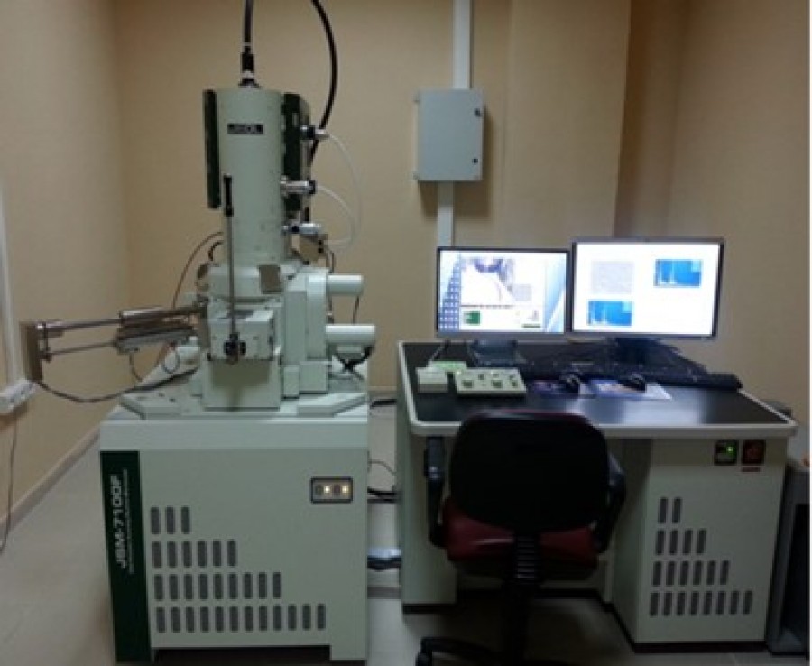 |
Working Principle:
SEM (Scanning Electron Microscope) is a type of electron microscope that focuses a beam of electrons onto the surface of a sample to obtain an image from that surface. Electrons interact with the atoms of the sample, producing various signals. The detection of these signals provides information about the surface topography and composition of the sample. The device includes an EDX (Energy Dispersive X-ray Spectrometer) detector that provides atomic percentages of elements in the sample. Mapping studies are also conducted. |
||
| Applications: SEM is widely used in various fields such as materials science, chemistry, pharmacy, forensic science, geology, dentistry, and the construction industry. With this device, it is possible to obtain information about the morphology, size, and composition properties of ceramics, metals, polymers, thin films, geological materials, and biological samples. Sample Acceptance Conditions: For powder samples, a minimum of 20mg is required. The sample size should not exceed 1cmx1cm. |
|||
| Instrument: TEM (Transmission Electron Microscope) Brand / Model: JEOL JEM-1400 PLUS | |||
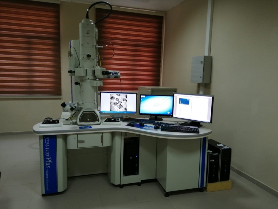 |
Working Principle: TEM (Transmission Electron Microscope) is based on the principle of imaging high-energy electrons passing through a very thin sample. This technique allows for obtaining morphological, structural, and elemental information at various magnifications, ranging from low to high. The device includes an EDX (Energy Dispersive X-ray Spectrometer) detector that provides atomic percentages of elements in the sample. |
||
| Applications: TEM (Transmission Electron Microscope) is used for various analyses in the fields of biology, medicine, and basic sciences. It allows for detailed, high-resolution, and high-contrast imaging of the internal structure of samples, as well as morphological studies and interpretation of structure-function relationships. Sample Acceptance Conditions: A minimum of 10 mg of powder sample should be provided. For powder samples, they are suspended in a suitable liquid in an Eppendorf tube and subjected to ultrasonication until homogeneously dispersed. Then, 5-10 ml of the suspension is dropped onto a carbon-coated grid and allowed to dry. |
|||
| Instrument: Atomic Force Microscope (AFM) Brand / Model: WITEC ALPHA 300RA | |||
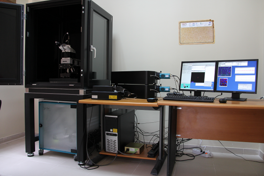 |
Working Principle:
Atomic Force Microscope (AFM) obtains three-dimensional topographic images of material surfaces by scanning the surface with a fine tip (cantilever). It is used to acquire various surface properties (surface morphology and roughness, etc.) of samples such as thin films, ceramics, metals, polymers, semiconductors, and biomaterials. AFM can be used in two different modes: the contact mode, where the tip makes contact with the surface, and the non-contact mode, where the tip does not touch the surface. It can also work in conjunction with Raman Spectroscopy, allowing for spectrum collection from the same region. |
||
| Applications: AFM can be applied in various fields such as solid-state physics, semiconductor technology, molecular engineering, polymer physics and chemistry, surface chemistry, molecular biology, and medicine. Sample Acceptance Conditions: Sample Acceptance Criteria: The minimum sample size requirement is a 1x1 cm sample in a coated form. |
|||